What is PSMA Imaging?
This video is an overview of PSMA-targeted PET imaging for prostate cancer. It explains how the scan works, when it is used, and how it helps doctors create more precise, personalized care plans.
Read the full video transcript below:
This video is an overview of PSMA-targeted imaging, also known as Prostate-Specific Membrane Antigen targeted imaging, and why it matters in prostate cancer care.
When diagnosing and monitoring prostate cancer, doctors rely on tests like PSA (Prostate-Specific Antigen) blood tests, biopsies, and conventional imaging scans such as CT or MRI. While these tests provide valuable information about the prostate cancer size and location, they don’t always detect small or hidden tumors with high accuracy.
That’s where PSMA-targeted PET imaging comes in.
What is PSMA Imaging, and how does it work?
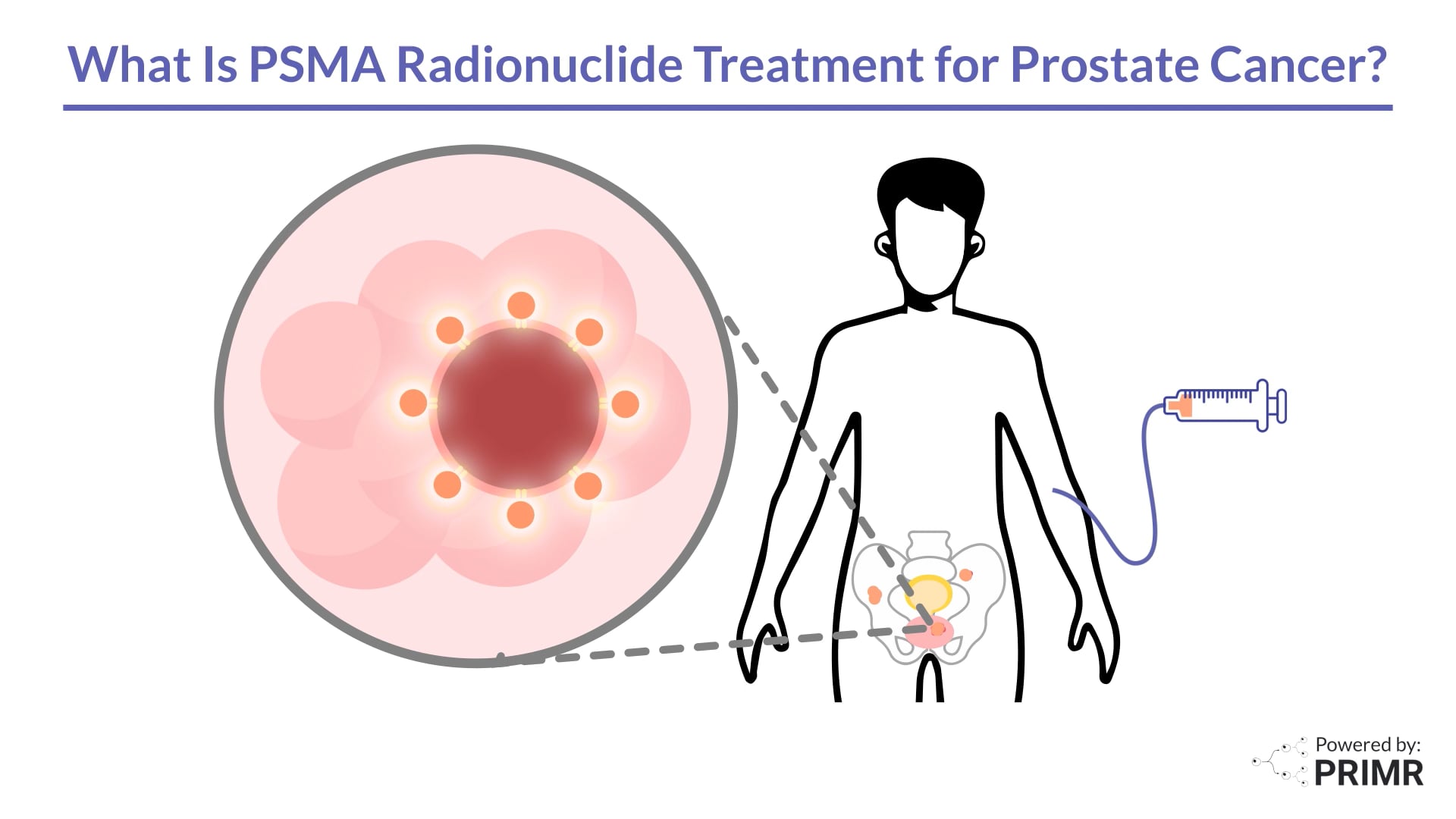
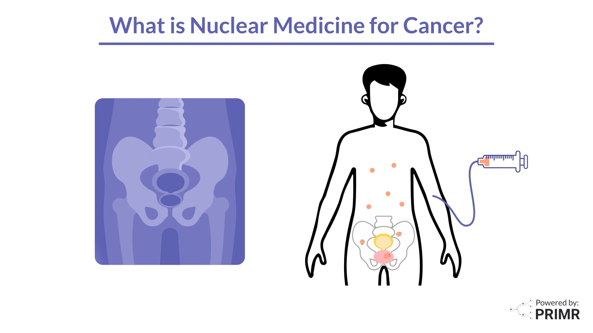
A PET scan, which stands for Positron Emission Tomography scan, is a medical imaging test that creates a 3D image of a patient’s internal organs and tissues. A liquid radioactive drug, sometimes referred to as a “radiopharmaceutical”, is injected into the patient prior to the PET scan and can highlight cancerous cells on the image. For some suspected prostate cancer, a radioactive isotope is attached to a PSMA-targeting molecule and then injected into a patient. Patients then undergo a PET scan where their internal organs can be seen via a PET camera. This allows doctors to see the tumors.
The PSMA imaging process involves three key steps.
First, a patient is injected with radioactive medicine that is designed to attach to PSMA proteins which appear on the surface of prostate cancer cells. There are multiple products designed to do this each having unique properties. One key difference between different radioactive medication is half-life. Half-life refers to the time it takes for half of the atoms that are radioactive to decay. Half-life is different for each radioisotope and is crucial in determining how long a radioactive substance will remain active within a body.
Next, after a short waiting period that can be unique to the radiopharmaceutical injected, the patient undergoes a PET scan, often combined with a CT or MRI. The scan detects the small amount of radioactivity from the tracer in your body and uses it to create images. In a PSMA PET scan, the images show where prostate cancer is in the body, because the medicine attaches to a protein found on prostate cancer cells. The CT or MRI portion of the scan shows the structural parts in the body.
Finally, specially trained doctors carefully review the results of the scan. They look for signs of cancer and determine where it is in the body. The information from a PSMA PET scan helps guide the development of a treatment plan. Since every patient is different, doctors will recommend treatment options based on whether PSMA-positive prostate cancer is found, how large any tumors are, and if and where it has spread.
How is PSMA Imaging Used in Prostate Cancer Care?
Doctors use PSMA PET imaging at different stages of prostate cancer diagnosis and treatment.
For initial diagnosis and staging, a PSMA PET scan can reveal whether the cancer is limited to the prostate or has spread to other areas often referred to as “metastatic prostate cancer”.
When it comes to treatment planning, PSMA PET imaging helps doctors choose the best treatment, such as surgery, radiation, non-radioactive drug therapy, or PSMA-targeted therapy referred to as RLT (Radioligand Therapy).
If a patient’s PSA levels start to rise after treatment, this can be referred to as “Biochemical Recurrence” or BCR for short. PSMA imaging can help detect the recurrent cancer, guiding further therapy.
Doctors will use physical exams, various blood tests, and/or imaging scans to monitor treatment.
Benefits of PSMA Imaging
PSMA imaging offers high accuracy, helping doctors find cancer. It also allows for staging, giving a picture of how far the cancer has spread. With this information, doctors can create a personalized treatment plan, targeting the cancer with the right therapies. And PSMA imaging may help avoid unnecessary procedures.
Who Can Benefit from PSMA Imaging?
PSMA PET scans are typically recommended for:
- Patients with newly diagnosed prostate cancer who are in intermediate or high-risk groups.
- Men with rising PSA levels after treatment, as it can help detect if the cancer has recurred or come back.
- Patients considering targeted therapies that depend on PSMA expression.
Ultimately, your doctor will determine if PSMA imaging is the right choice for you based on your specific case and medical history.
This is not medical advice. This video is for educational purposes only. Talk to your doctor before making any medical decisions.

.jpg)
.jpg)
%20Thumbnail.png)


.jpg)
.png)


.jpeg)


.webp)


.jpeg)

