Understanding Dosimetry in PSMA Radioligand Therapy
Radioligand therapy, or RLT, is a targeted treatment approach for prostate cancer that delivers radiation directly to PSMA-avid cancer cells. This video explains dosimetry, how absorbed radiation dose is measured after PSMA radioligand therapy, why dose to normal organs matters, and how imaging may help guide patient monitoring, toxicity risk assessment, and future personalized treatment planning.
Developed in collaboration with Curium Pharma.
Read the full video transcript below:
Radioligand therapy, or RLT, is changing how we treat advanced prostate cancer. As adoption grows, dosimetry (especially off-target dosimetry) is becoming more important to optimizing outcomes.
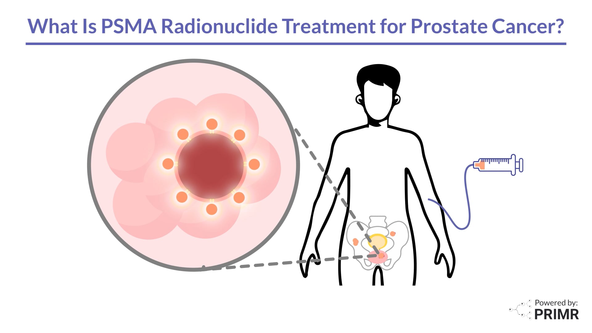
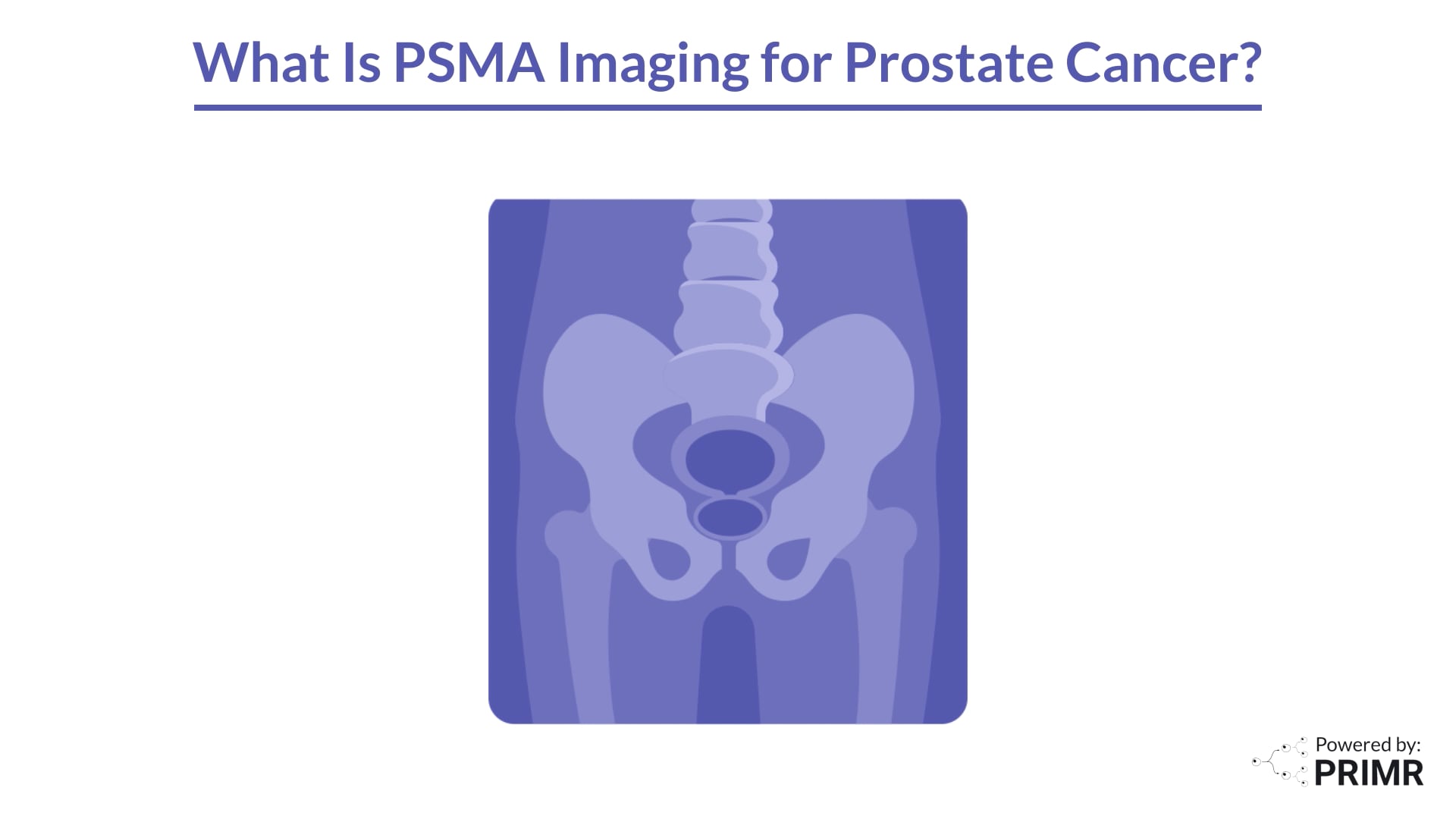
PSMA has long been an important marker in prostate cancer. PSMA-targeted radioligands are increasingly being used to help guide prostate cancer care decisions, inform prognosis and influence treatment selection, whether that’s surgery, radiation, systemic intensification or PSMA-targeted radioligand therapy. PSMA PET/CT imaging helps to define the disease extent and location by identification of PSMA-avid prostate cancer cells. PSMA radioligand therapy pairs a PSMA-targeting component with a radionuclide payload.
Activity localizes to the PSMA-avid tumor and contributes to tumor doses. This makes dosimetry more important. It helps us treat the tumor while protecting normal organs—and it helps us anticipate side effects.
Dosimetry is the quantification of absorbed radiation dose delivered to tissues after administration of a radiopharmaceutical, typically reported in units of Gray. In PSMA radioligand therapy, the intent is straightforward: the targeting molecule binds to PSMA on tumor cells, concentrating radioactivity in tumor sites. However, biodistribution is never perfectly tumor-selective. Dosimetry is how we describe where the radioactivity actually goes over time, and what that implies for both tumor and healthy tissues.
Conceptually and as previously stated, radioligand therapy pairs a PSMA-targeting component with a radionuclide payload. When activity localizes to a tumor, it contributes to tumor dose. When it does not, it contributes to normal-organ dose. Dosimetry helps quantify both, and it highlights inter-patient variability. Two patients can receive the same administered activity and experience meaningfully different absorbed doses to key organs.
In practice, dosimetry is often derived from post-therapy imaging.
SPECT/CT—single-photon emission computed tomography paired with CT—is commonly performed after a treatment cycle to map radioactivity distribution and estimate absorbed dose.
Dosimetry is often framed as “dose to the target.” In this video, the focus is on dose to healthy tissues, because those organs at risk can determine tolerability and may limit therapy over time.
Why does this matter? Radioligand therapy is a balancing act: maximizing tumor dose while limiting exposure to radiation-sensitive organs. Understanding distribution helps anticipate toxicity, supports patient monitoring, and, over time, may enable more individualized approaches.
After PSMA radioligand therapy, measurable exposure can occur in normal tissues, including salivary glands, lacrimal glands, kidneys, and bone marrow. There are two main drivers. One is uptake in normal tissues with physiologic PSMA expression. The other is systemic off-target exposure from circulating activity that isn’t bound to tumor at that time.
Off-target radiation becomes clinically relevant when evaluating baseline PSMA expression in healthy tissue to help predict toxicity risk.
For example, salivary gland uptake metrics on baseline PSMA imaging have been associated with xerostomia risk after therapy.
For hematologic toxicity, baseline disease burden—especially in bone—may be important. In one analysis, higher baseline bone Total Lesion PSMA, or TLP—a PET-derived measure that combines PSMA-avid tumor volume and average uptake—was associated with earlier onset of severe hematologic toxicity.
In a clinical trial evaluating a radioligand therapy plus standard of care versus standard of care alone, absorbed-dose analyses provide a framework for understanding organ exposure at scale. In reported analyses, patients who experienced renal toxicity had numerically higher absorbed doses.
The point is not that a single number perfectly predicts toxicity for every patient. The point is that dosimetry provides a measurable bridge between administered activity, biodistribution, and clinically meaningful adverse events, supporting better counseling, monitoring, and supportive care planning.
Dosimetry literacy benefits the entire care team, including nuclear medicine physicians, urologists, medical oncologists, and radiation oncologists. It supports therapy planning, frames downstream adverse-event risk, and helps teams anticipate the level of supportive care a given patient may require.
Ultimately, dosimetry is about balance: maximizing tumor control while managing the risk of off-target toxicity. Ongoing research will refine imaging-derived predictors and absorbed-dose relationships and may enable more personalized dosing strategies as evidence matures.
This educational video is intended for healthcare professionals and does not provide medical advice. Clinical decisions should be individualized and based on the full clinical context, local protocols, and approved prescribing information.
.jpg)
.jpg)
%20Thumbnail.png)


.jpg)
.png)
.png)
.jpg)

.jpg)
.png)


.jpeg)


.jpeg)

